Research Topics
Seeing, Hearing, and Feeling in the Nanoworld
Nanomechanical tissue properties will be made accessible to the human senses
 In collaboration with the Cognitive Systems Lab, we are developing a multisensory display that allows the user to perceive the nanomechanical properties of a sample, which we measure using atomic force microscopy, trough touch and other human senses. We take a multidisciplinary approach, with an emphasis on fundamental phenomena and the goal of developing exciting new applications in the life sciences. (Illustration: R. Magerle)
Collagen Fibrils
Nanomechanical properties of the most abundant structural protein in vertebrates
 Fibril-forming collagens are a major component of the connective tissues of vertebrates. The amount of water molecules and the type and distribution of molecular cross-links between the collagen molecules decisively determine the mechanical properties of collagen fibrils. We study collagen fibrils using atomic force microscopy, whereby we can tune the water content of the fibrils very accurately via the relative humidity. (Figure: R. Magerle, M. Dehnert, D. Voigt, A. Bernstein, Analytical Chemistry 92, 8741–8749 (2020); © 2020 American Chemical Society.)
EPICAL Hysteresis Model
Rate-independent energy dissipation in collagen fibrils
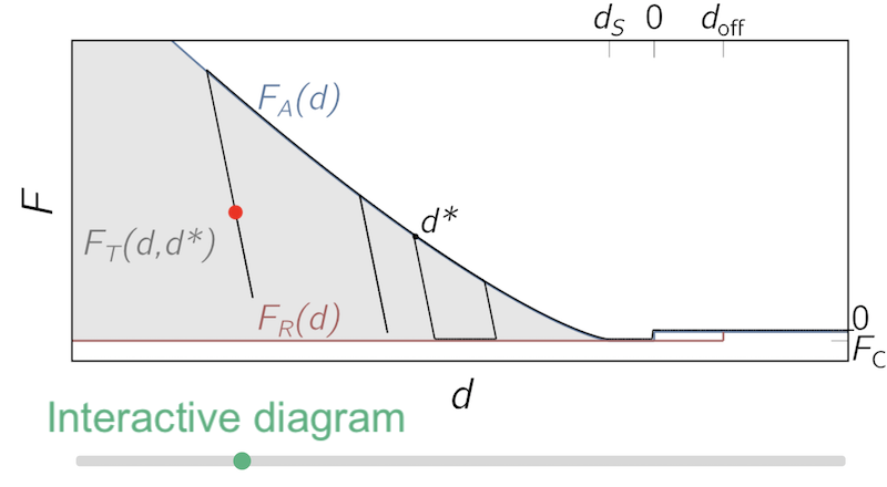 The EPICAL hysteresis model describes phenomenologically and in a unified form the sequences of mechanical phenomena that occur in AFM-based nanoindentation experiments: Elasto-plastic indentation, capillary adhesion and surface levelling. The model is based on force-distance data measured with the atomic force microscope (AFM) during a large approach-retract cycle and predicts the force (output) and the dissipated energy for arbitrary indentation trajectories (input). (Figure adoped from: R. Magerle, P. Zech, M. Dehnert, A. Bendixen, A. Otto, Soft Matter 20, 2831–2839 (2024); © 2024 The Royal Society of Chemistry)
MUSIC-Mode Atomic Force Microscopy
Nanoscale depth profiles of soft polymer surfaces and fluids
 In tapping mode AFM, we can measure the indentation depth of the tip into the sample and use this information to reconstruct spatial depth profiles of the sample surface, e.g. of polymer-functionalized graphene oxide layers (see figure) as well as of semi-crystalline polymers and collagen fibrils. A variation is the multi-set point intermittent contact (MUSIC) mode, which can be used to image soft nanoscale objects and even liquids without the artifacts of feedback control. (Figure: M. Dehnert, E.-C. Spitzner, F. Beckert, C. Friedrich, R. Magerle, Macromolecules 49, 7415–7425 (2016); © 2016 American Chemical Society.)